
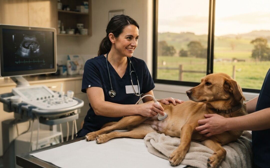
A pet ultrasound can show the size and texture of organs, soft-tissue changes, masses and fluid inside the body. Knowing what a vet can see on a pet ultrasound helps explain why vets use it to investigate concerns such as abnormal test results, vomiting or weight loss, and why they may pair it with other diagnostic tests.
It’s understandable to want a clearer picture of what a scan might reveal when your pet isn’t feeling well. Ultrasound uses sound waves, not radiation, to create images of organs and soft tissues. It can also show some structures in motion, including the heart as it beats.
This guide explains how pet ultrasounds work, what vets may examine and how the images can help investigate health concerns. You’ll also learn what the procedure may involve and how ultrasound’s strengths and limits shape the information it provides.
Key Takeaways
- Ultrasound helps vets examine soft tissues and organs, while X-rays are generally better for assessing bones.
- A scan may help investigate changes in organs, fluid build-up or a soft-tissue mass, but its usefulness depends on the area being examined.
- Ultrasound images can show movement, helping vets assess structures such as the heart and monitor pregnancy.
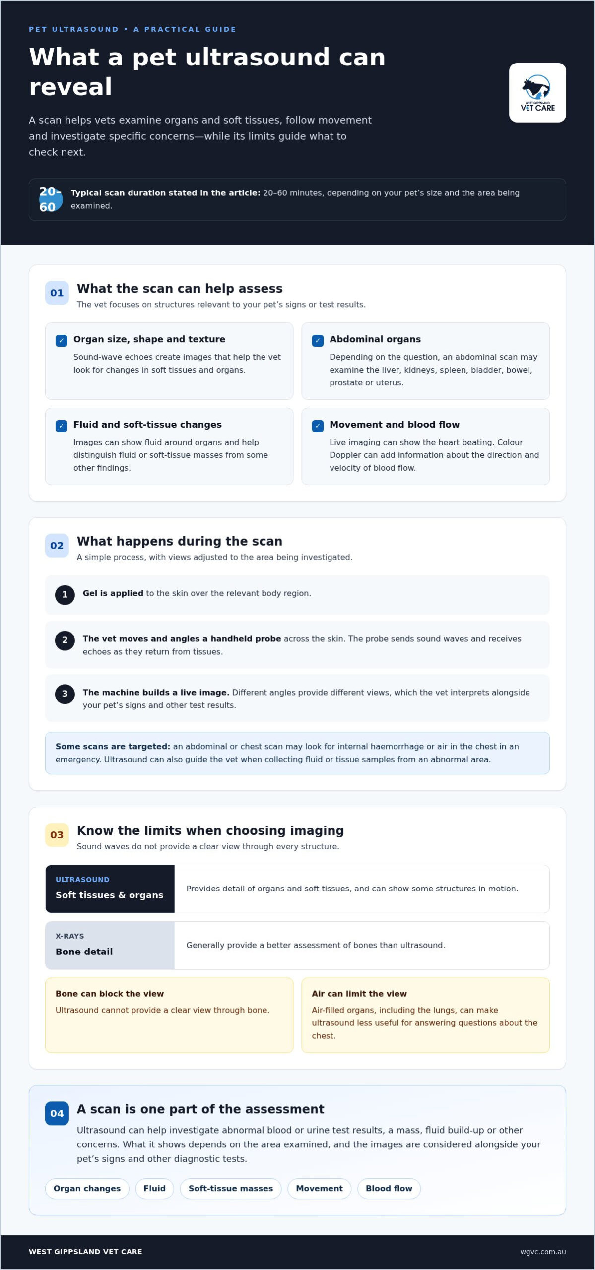
- Most pet ultrasounds take 20-60 minutes, and the procedure usually involves gel and a handheld scanner moved over the skin.
- Knowing what ultrasound can and can’t show can help you understand why a vet may use it alongside other diagnostic tests.
What Happens During a Pet Ultrasound?
A pet ultrasound uses sound waves to create images of internal organs, soft tissues and fluid-filled spaces. The vet moves a handheld probe over the skin to examine the area relevant to your pet’s signs or test results.
Soft-tissue and organ assessment
Ultrasound examines soft tissues by sending sound waves into the body and turning their returning echoes into images. This matters because vets can assess an organ’s size, shape and texture, then look for changes such as a mass or fluid. For example, an abdominal scan can help investigate unusual blood or urine test results by showing several organs in one examination.
Ultrasound limits around bone and air
Ultrasound is less effective when sound waves meet bone or air, so it cannot provide a clear view through these structures. This matters when choosing an imaging method: X-rays generally show bones better, while ultrasound provides more detail of soft tissues and organs. Air-filled organs, including the lungs, can also limit the view, so a scan may not answer every question about the chest.
Visible organs and structures
An abdominal ultrasound can show the liver, kidneys, spleen, urinary bladder, bowel, prostate or uterus. The vet focuses on structures relevant to the concern, such as checking the bladder for stones or examining the kidneys after an abnormal urine result. Ultrasound can also show fluid around organs and help distinguish fluid or soft-tissue masses from some other findings.
The scan usually takes 20-60 minutes, depending on your pet’s size and the area being examined. The vet applies gel to the skin, then moves the probe across the relevant body region. Each angle gives a different view, allowing the vet to assess internal structures rather than relying on a single image.
Some examinations focus on a specific question. An abdominal scan may include the lymph nodes, spleen, kidneys, liver and bladder when test results are abnormal. In an emergency, a focused scan of the abdomen or chest can look for internal haemorrhage or air in the chest. These targeted examinations help guide what the vet investigates next.
Ultrasound can also guide a vet collecting fluid or tissue samples from an abnormal area. The images help locate a mass or fluid pocket during sampling. A sample can provide information for diagnosis and treatment planning, while the images themselves are only one part of assessing your pet’s health.
How does an ultrasound work?
Veterinary ultrasound turns sound waves into images that help examine an animal’s internal organs. A handheld probe sends sound waves into the body, then receives echoes as they bounce back from tissues. The machine processes those echoes into a live image on its screen. The vet moves and angles the probe to view the area from different directions.
Different tissues return different echoes, creating contrasts between structures. Fluid-filled areas often appear dark, while tissue boundaries and denser structures create brighter patterns. These differences help the vet assess an organ’s shape and texture. For example, an abdominal scan can show the bladder and nearby tissues together, helping the vet examine their position and appearance.
From sound waves to a live image
The probe both sends and receives sound waves. As the vet moves it across the skin, each position creates another view of the structures below. The machine updates the image as the probe changes angle, allowing the vet to follow an organ across several views rather than relying on a single still picture.
Live imaging can show movement inside the body. The heart, for example, can be watched as it beats, allowing the vet to assess its structure and function in motion. Images may show how the heart chambers and valves behave during a heartbeat. This moving view adds information that a still image alone cannot provide.
Seeing blood flow and the heart
Some ultrasound machines can display blood flow as well as organ structures. Colour Doppler ultrasound measures the direction and velocity of blood moving through the heart. This helps the vet assess how blood travels through cardiac structures, alongside the heart’s movement. The colour display represents flow information, not a photograph of the blood itself.
These views help answer different clinical questions. A standard image can show the heart’s shape and movement, while Doppler adds information about blood flow. The vet selects the view that matches the concern, such as assessing heart function or investigating an abnormal finding. Each view contributes a different part of the examination.
Ultrasound imaging also depends on where sound waves can travel and return. Air and bone interfere with clear images, so ultrasound is less useful for examining air-filled lungs or viewing bone detail. X-rays usually provide a clearer assessment of bones. Knowing these limits helps explain why a vet may use different imaging methods for different body areas.
For an abdominal examination, the vet guides the probe over the area being assessed and adjusts its angle to inspect internal structures. The screen updates as the probe moves, allowing the vet to compare tissue boundaries and organ appearances during the scan. The vet interprets these images alongside your pet’s signs and other test results.
How can an ultrasound help my pet?
A reproductive ultrasound uses sound waves to examine pregnancy and reproductive organs in dogs and cats. It can help assess pregnancy progress, foetal health and possible complications.
Pregnancy and foetal monitoring
For dogs and cats, pregnancy ultrasounds are typically performed around 25-35 days after mating. The images can help assess whether pregnancy is progressing and show developing foetuses. This gives the vet information about foetal health and potential complications, supporting decisions about care during pregnancy.
Ultrasound can also examine reproductive organs beyond pregnancy. A reproductive scan may assess the uterus, ovaries or prostate for changes such as ovarian cysts or prostatitis. By showing internal structures, the scan helps the vet evaluate reproductive health beyond outward signs. It is a painless way to gather information about these organs.
Reasons a vet may recommend ultrasound
A vet may recommend an ultrasound to assess a pregnancy or investigate suspected reproductive disease. Signs such as vomiting, diarrhoea or unexplained weight loss can also prompt investigation, particularly when other findings point to an internal concern. Changes in blood or urine test results may lead to an abdominal scan of organs such as the kidneys.
The reason for imaging determines which area the vet examines. For pregnancy, the vet focuses on the uterus and developing foetuses. For other concerns, an abdominal scan can assess organs including the kidneys, liver, bladder and spleen. Ultrasound can help investigate changes in these structures and identify findings that need further assessment.
Pregnancy monitoring is one part of a broader diagnostic process. Ultrasound findings can be considered alongside your pet’s history, physical examination and test results. For example, a scan may provide information about reproductive organs while the vet also considers symptoms or abnormal results. Together, these details help build a clearer picture of your pet’s health and possible next steps.
If you’re planning a litter, the timing of the scan matters. The typical window of 25-35 days after mating gives the vet a chance to assess pregnancy and foetal development. Tell the vet the mating date and any changes you’ve noticed, such as altered appetite or behaviour. Those details help place the ultrasound findings in context.
Ultrasound can support monitoring, but it doesn’t replace a full veterinary assessment. Whether the concern is pregnancy, reproductive health or symptoms such as vomiting, the images help the vet investigate internal changes and decide what information is needed next.
Safety and comfort during the procedure
Most routine pet ultrasounds can be performed without sedation or anaesthesia. The scan itself doesn’t use radiation; risks are associated with sedation, anaesthesia or collecting tissue or fluid samples, rather than ultrasound technology. A scan usually takes 20-60 minutes, depending on your pet’s size and the area examined.
Preparation depends on the body area being scanned. For a planned ultrasound, a dog may need to fast for a few hours. If possible, dogs and cats should avoid urinating for about three to six hours beforehand, so the bladder is full enough to examine. Follow the vet’s instructions, especially for exotic pets, as fasting advice varies by species.
During the procedure, the vet applies gel to the skin and moves a handheld transducer across the area. The gel helps the probe make contact, while the screen displays images of internal organs and soft tissues. The process is usually painless, and pets generally tolerate it well. For an abdominal scan, the vet may examine structures such as the spleen and bladder from different angles.
Sedation may be recommended if a pet is aggressive, too anxious or painful to remain still, or is having an ultrasound-guided tissue biopsy. The vet weighs the need to keep the animal still against the risks associated with sedation or anaesthesia. When needed, these medicines, rather than the sound waves, are the source of ultrasound-related procedural risk.
Ultrasound can guide fluid or tissue sampling when an image shows an area that needs closer investigation. For example, if the vet finds an abdominal mass or abnormal fluid build-up, they may recommend collecting a sample. Ultrasound-guided biopsy can be less invasive than surgery for obtaining tissue, although sampling has risks separate from the scan.
Most pets don’t need recovery time after a routine ultrasound. If sedation or anaesthesia is used, recovery relates to those medicines and your pet’s circumstances, rather than the imaging itself. A calm, still position helps the vet capture clear images, so the team may adjust your pet’s position during the scan.
When a radiologist needs to interpret the images, results may take a day or two. The time to receive findings therefore depends partly on whether specialist interpretation is needed. Your vet can explain what the images show and how they fit with your pet’s examination or other test results.
Conditions ultrasound may help diagnose
Ultrasound can help investigate abnormalities in the abdomen, chest, heart and soft tissues. The vet chooses the area based on your pet’s signs, examination and other test results.
Heart conditions
A cardiac ultrasound examines the heart’s structure and function, including its movement as it beats. It can help identify heart conditions such as mitral valve disease in dogs or hypertrophic cardiomyopathy in cats. Some machines also show blood flow, giving the vet another way to assess how the heart is working. The images are interpreted alongside your pet’s clinical signs.
For abdominal concerns, ultrasound may help identify gastrointestinal disease or blockage, inflammation, bladder stones, tumours and fluid build-up. It can also examine organs such as the pancreas, liver, kidneys, spleen and intestines. For example, a scan may help investigate vomiting, diarrhoea or weight loss, or look for internal bleeding when other findings raise concern.
Cancer and mass assessment
Ultrasound can detect a mass and show details such as its size, location and appearance. These features may help a vet distinguish a mass from fluid or another soft-tissue finding, and contribute to assessing whether it appears benign or malignant. However, an image alone cannot confirm cancer or reliably determine a mass’s nature.
If a mass or abnormal fluid is found, the vet may recommend collecting a sample for further examination. Ultrasound can guide sampling by helping locate the area of concern. The sample provides information that imaging alone cannot and may help guide decisions about further investigation or treatment.
Ultrasound can also assess muscles, tendons, ligaments and joints when a pet has lameness or pain. This can help investigate soft-tissue injuries and inflammatory conditions. It is a different diagnostic use from examining abdominal organs, and the area being scanned shapes what the images can reveal.
The sound waves used for ultrasound create images of internal structures, but the scan has limits. Bone and air can obstruct the view, so ultrasound is less suited to assessing bones or air-filled lungs. X-rays may provide clearer bone detail, while ultrasound can show soft-tissue findings, fluid and organ changes that help build a fuller diagnostic picture.
For concerns such as a new mass, persistent vomiting or suspected heart disease, diagnostic imaging can help clarify what is happening inside your pet’s body. Discuss diagnostic imaging for your pet as part of understanding the next steps in their care.
West Gippsland Vet Care: Pet Vaccinations
West Gippsland Vet Care supports your pet’s ongoing health with pet vaccinations and other routine care. After investigating a health concern, you can also discuss preventive care or a separate procedure suited to your pet’s needs.
Vaccinations help you plan for your pet’s health, while desexing procedures and microchipping address other important aspects of care. For pets with mobility concerns or orthopaedic conditions, orthopaedic surgery may be part of the treatment discussion. Each service serves a different purpose, so the next step depends on your pet’s needs.
Contact West Gippsland Vet Care to discuss your pet’s care and the questions you’d like answered.
Make a clear plan for your pet’s care
Understanding what a vet can see on a pet ultrasound is a useful starting point, but the next step is connecting those images with your pet’s symptoms and overall health. Note any changes you’ve noticed and the questions you want answered. A clear conversation about findings and follow-up can help you feel prepared to make decisions for your companion.
West Gippsland Vet Care can support your pet beyond an investigation with care such as pet vaccinations, desexing procedures and microchipping. These services address different needs, from preventive health to identification, and can be discussed as part of your pet’s ongoing care.
Contact West Gippsland Vet Care today to talk about your pet’s needs and the care options available. Taking the next step can feel easier when you have a trusted team to talk things through with.
Frequently Asked Questions
What Is an Ultrasound for Pets?
A pet ultrasound is a veterinary imaging examination that helps investigate a specific health question inside your animal’s body. For example, a vet may focus on the abdomen after unusual blood or urine test results rather than scanning every area. The images can guide further investigation, such as deciding whether a mass or fluid build-up needs to be sampled for closer examination.
Does an ultrasound hurt my pet?
A routine ultrasound is usually painless. The vet applies gel and moves a probe over your pet’s skin. It may feel unfamiliar, but it does not involve cutting or injections. Your pet may need to hold a position while images are taken. If an area is painful or your pet is very anxious, the vet considers comfort and stillness when planning the examination.
Is a pet ultrasound safe?
Ultrasound uses sound waves, not radiation, and the scan itself is considered safe. Any risks are associated with sedation, anaesthesia or collecting tissue or fluid samples, rather than the ultrasound technology. Most routine scans don’t require sedation. If your pet needs a guided biopsy, the vet weighs the reason for sampling and the associated procedure risks when planning care.
What Can an Ultrasound Diagnose?
An ultrasound can help investigate concerns such as gastrointestinal disease, blockages, bladder stones, organ abnormalities and heart disease. It gives the vet information about internal structures to support diagnosis alongside an examination and other test results. For instance, a scan may help explore persistent vomiting or weight loss. The images guide further investigation, but a mass’s appearance alone doesn’t confirm its cause.
What can a pet ultrasound detect?
A pet ultrasound can reveal findings such as fluid in the abdomen or chest, cysts, masses, internal bleeding and changes in organ size or texture. It can also help distinguish fluid or a soft-tissue mass from other findings. What a vet can see on a pet ultrasound depends on the area examined and whether air or bone obstructs the view.
What organs and tissues can a veterinarian examine on a pet ultrasound?
A vet can examine structures including the liver, kidneys, spleen, bowel, bladder, prostate and uterus. The heart can also be assessed, including its movement as it beats. The area chosen depends on the reason for imaging. For example, an abdominal scan may include the bladder and kidneys when urine test results are abnormal, while a cardiac scan focuses on heart structure and function.
What conditions can an abdominal ultrasound detect in dogs and cats?
An abdominal ultrasound can help investigate blockages, tumours, inflammation, chronic bowel disease and bladder stones in dogs and cats. It can also show fluid build-up or organ changes involving structures such as the liver, pancreas, kidneys and spleen. Vets may use it when symptoms such as vomiting, diarrhoea or weight loss need investigation, or when blood and urine results show abnormalities.
Can a pet ultrasound show pregnancy, puppies or kittens, and fetal heartbeats?
An ultrasound can help assess pregnancy progress, developing puppies or kittens, and foetal health. For dogs and cats, pregnancy scans are typically performed around 25-35 days after mating. Ultrasound imaging can show structures in motion, including a heart as it beats. A scan also helps assess reproductive organs and potential complications, giving the vet useful information about pregnancy and the developing litter.